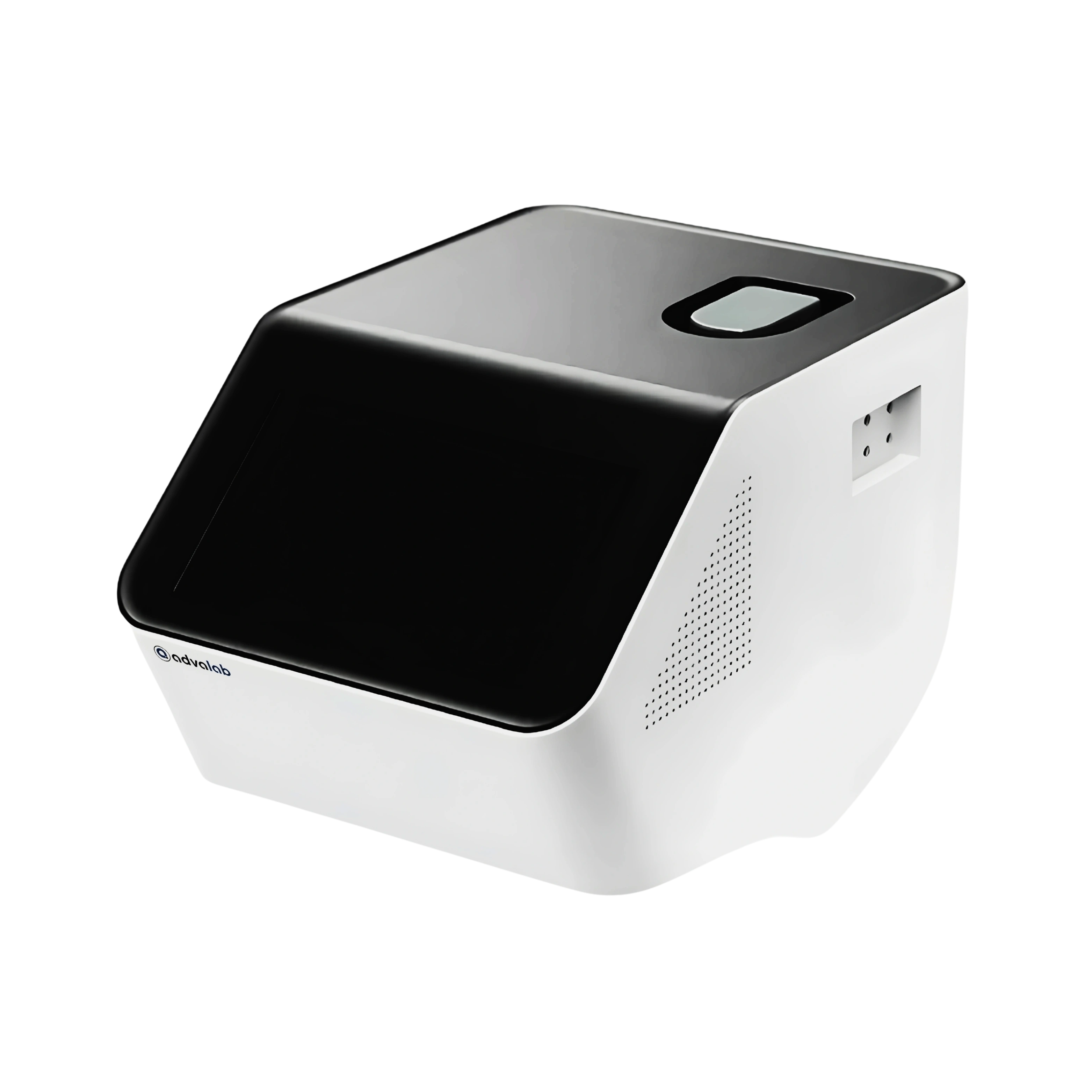
Cell Imaging Analyzer ADCIA-501 uses a sample volume of 10µl for rapid cell analysis while minimizing sample usage. It supports high-throughput screening with multi-well plate compatibility for large-scale experiments. The analyzer features auto-focus, image time-lapse imaging for improved data quality. This unit minimizes user intervention through automated workflows and intuitive software. Our Cell Imaging Analyzer is used in drug discovery, cancer research and immunology to visualize cellular behaviour.
| Sample Volume | 30 µl |
|---|---|
| Sample Concentration | 2×10⁴ to 1×10⁷cells/mL |
| Counting principle | Flow image method |
| Dual Detection Channels | 620 ± 30 nm / 520 ± 25 nm |
| Detection of Particle Size | 3 to 40 µm |
| Optical Channel | Brightfield image acquisition |
| Fluorescent Channel | 485 nm |
| Detection Time | < 30 sec |
Our Cell Imaging Analyzer ADCIA-501 is used for high-throughput, quantitative analysis of cellular structures and functions in drug discovery, cancer research and toxicity screening.