
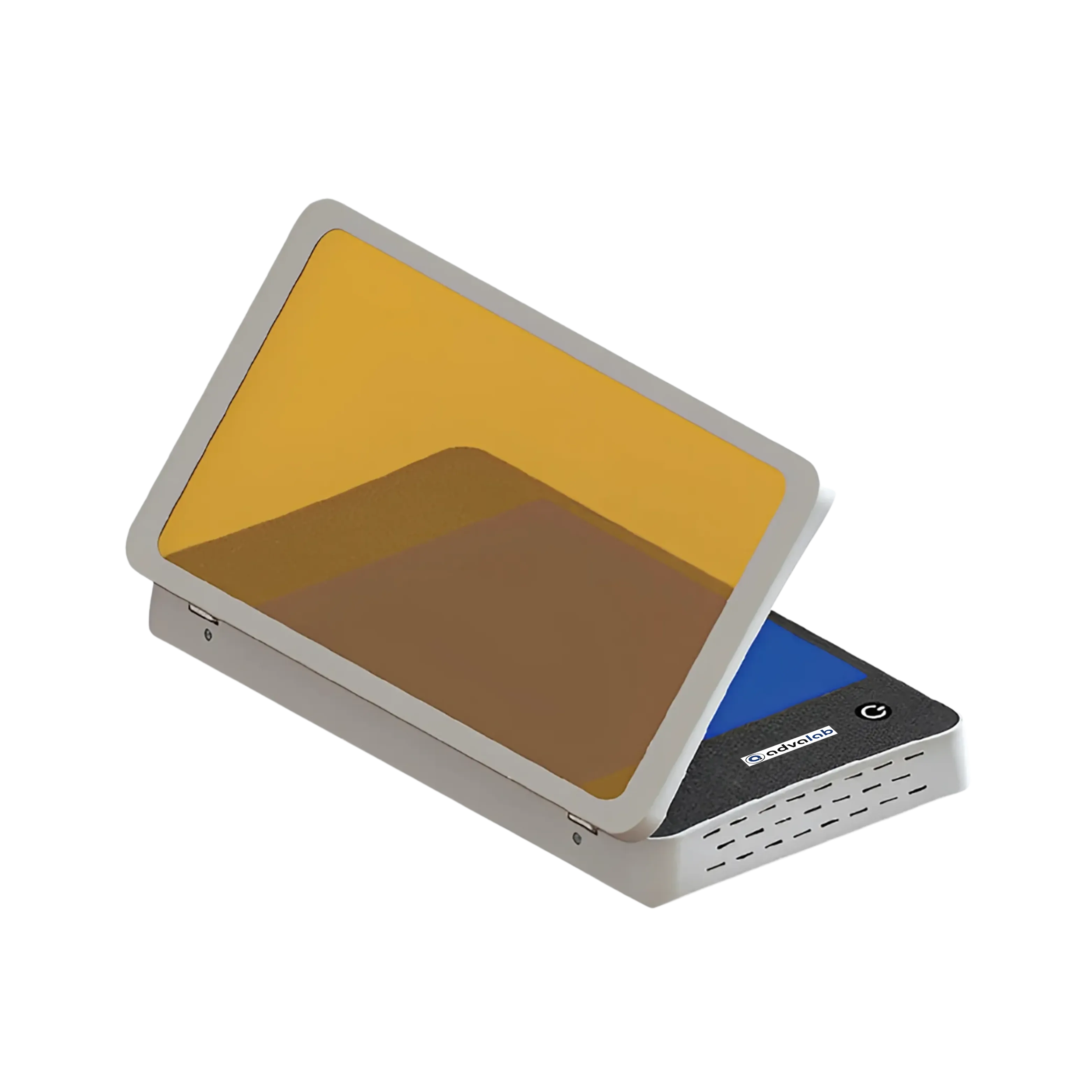
| Source UV wavelength (nm) | 470 nm |
| Transmission size (W×L) | 200×150 mm |
| Dimensions (L×W×H) | 310×180×50 mm |
| Weight (kg) | 1.5 kg |
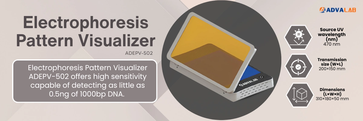
Our Electrophoresis Pattern Visualizer ADEPV-502 is widely used for observing, photographing and cutting DNA, RNA or protein electrophoresis gels stained with safe fluorescent dyes which makes it ideal for routine laboratory use.
1. What is the Electrophoresis Pattern Visualizer ADEPV-502 used for?
The Electrophoresis Pattern Visualizer ADEPV-502 is used to observe and document DNA, RNA, and protein samples in stained agarose or polyacrylamide gels. It uses blue LED light instead of UV, ensuring user and sample safety. This makes it ideal for research and educational labs focused on safe, eco-friendly practices. Its design supports easy imaging, gel cutting, and real-time observation.
2. Is the Electrophoresis Pattern Visualizer ADEPV-502 suitable for gel cutting or DNA band excision?
Yes, the Electrophoresis Pattern Visualizer ADEPV-502 is specifically designed for gel cutting and DNA band excision workflows. Its bottom-up illumination and adjustable filter allow clear visualization without glare. This ensures precise and efficient DNA band excision for downstream work. The flat surface also makes handling gels safer and easier.
3. How should I clean and maintain the Electrophoresis Pattern Visualizer ADEPV-502?
The Electrophoresis Pattern Visualiser ADEPV-502 requires minimal maintenance due to its solid-state LED design. Clean the surface with a soft cloth and mild detergent. Avoid abrasive cleaners to protect the optical filter. Store in a dry, dust-free area when not in use.
4. How sensitive is the Electrophoresis Pattern Visualizer ADEPV-502?
The Electrophoresis Pattern Visualizer ADEPV-502 can detect as little as 0.5ng of 1000bp DNA stained with efficient dyes. This makes it suitable for routine visualization and semi-quantitative analysis. Uniform LED light ensures sharp bands and low background. It supports both research and diagnostic use.
5. What are the advantages of three-stage dimming feature in the Electrophoresis Pattern Visualizer ADEPV-502?
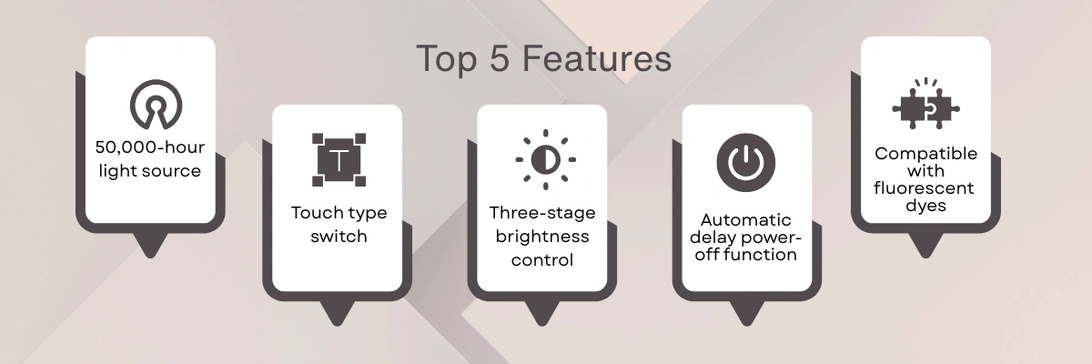
Three-stage dimming in the Electrophoresis Pattern Visualizer ADEPV-502 lets users match light intensity to stain levels and gel thickness. It reduces background in dense gels and enhances visibility in faint ones. This is crucial for sensitive tasks like CRISPR genotyping. It also lowers eye strain and extends LED life.
6. What is the purpose of an Electrophoresis Pattern Visualizer in the lab?
An Electrophoresis Pattern Visualizer is used to observe and analyze band patterns formed during gel electrophoresis. It helps researchers identify the presence, size, and intensity of DNA, RNA, or protein samples. Clear visualization enhances accuracy in molecular diagnostics and research. These tools are commonly used in biology, forensic, and clinical laboratories.
7. Are Electrophoresis Pattern Visualizers suitable for teaching labs?
These visualizers are well-suited for educational environments due to their simplicity and safety. They help students understand electrophoresis techniques through real-time observation of gel results. The systems are easy to operate, requiring minimal training. Their reliability makes them ideal for repeated classroom use.
8. What factors should be considered when choosing a Pattern Visualizer?
Important factors include the size of the viewing area, type of light source, and dye compatibility. Portability and ease of maintenance are also valuable for dynamic lab environments. The presence of brightness controls or filter options can improve result clarity. Always ensure the system meets safety standards and lab-specific requirements.
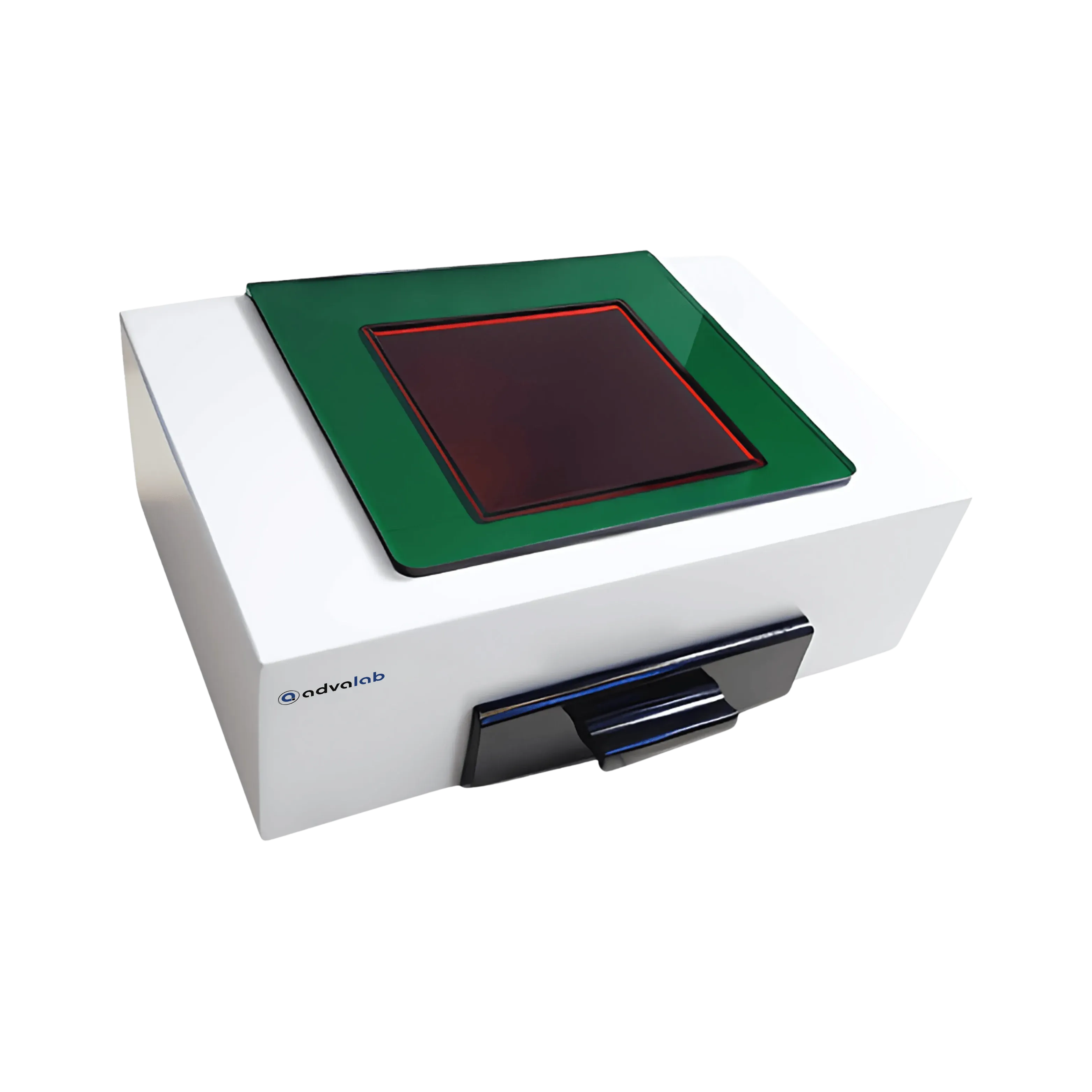
| Transmission size (W×L) | 120×120 mm |
| Dimensions (L×W×H) | 300×200×120 mm |
| Light Source | Visible Light (no UV) |